 |
 |
 |
 |
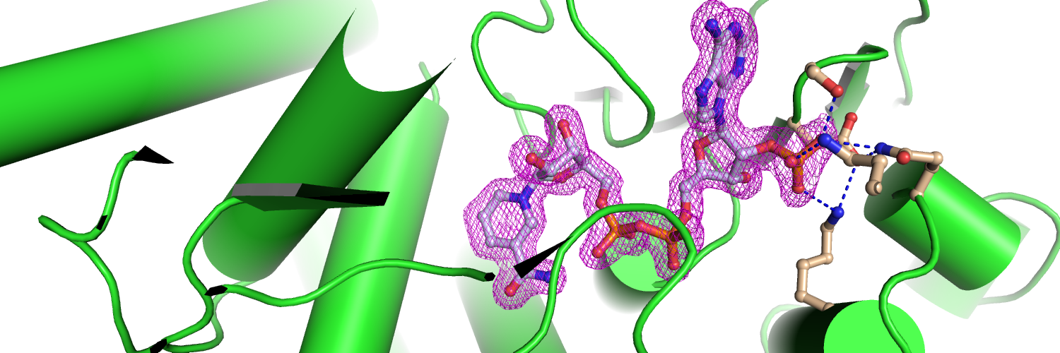
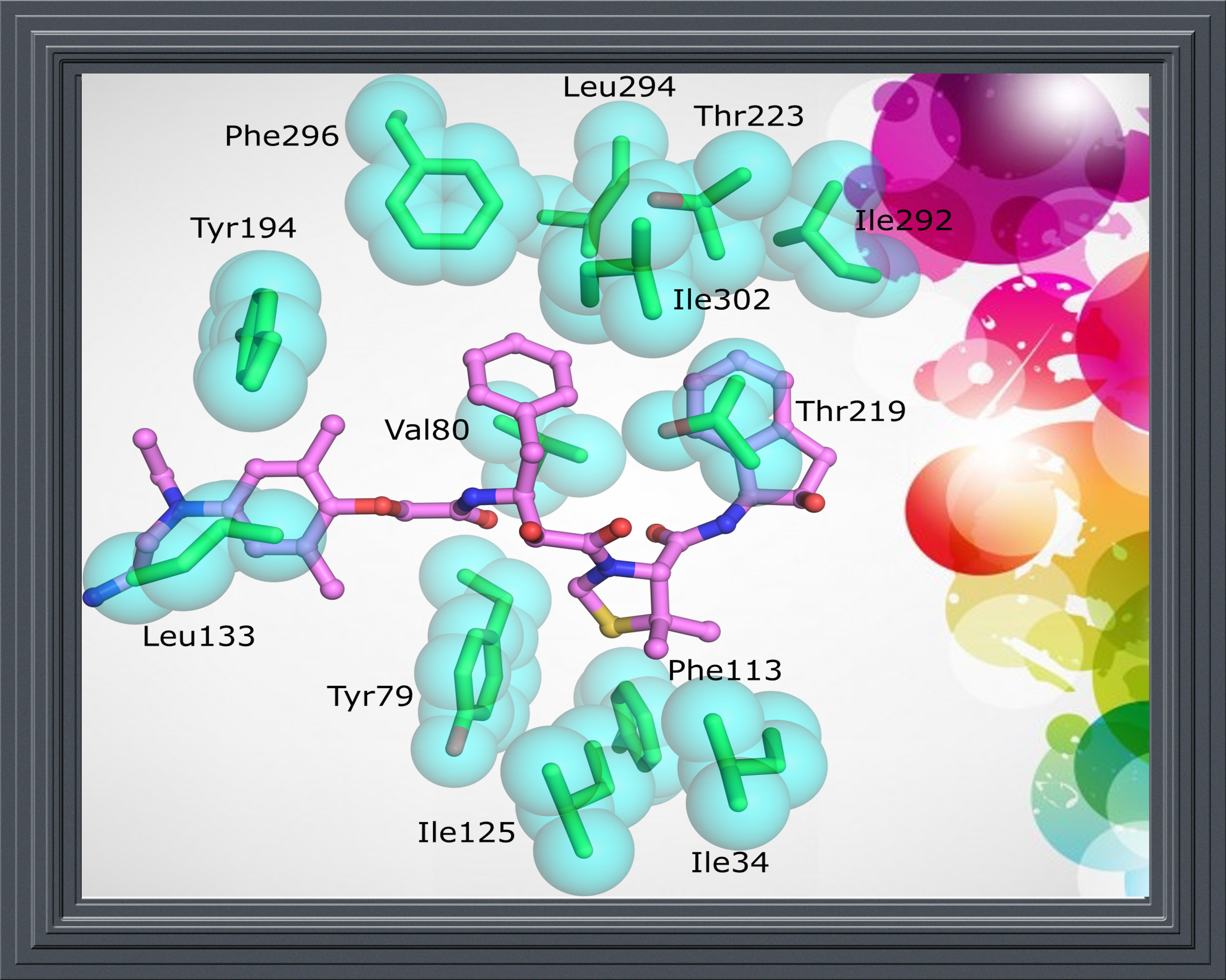
Crystal structure of plasmepsin II complexed with KNI-10333. PDB ID: 5YIC |
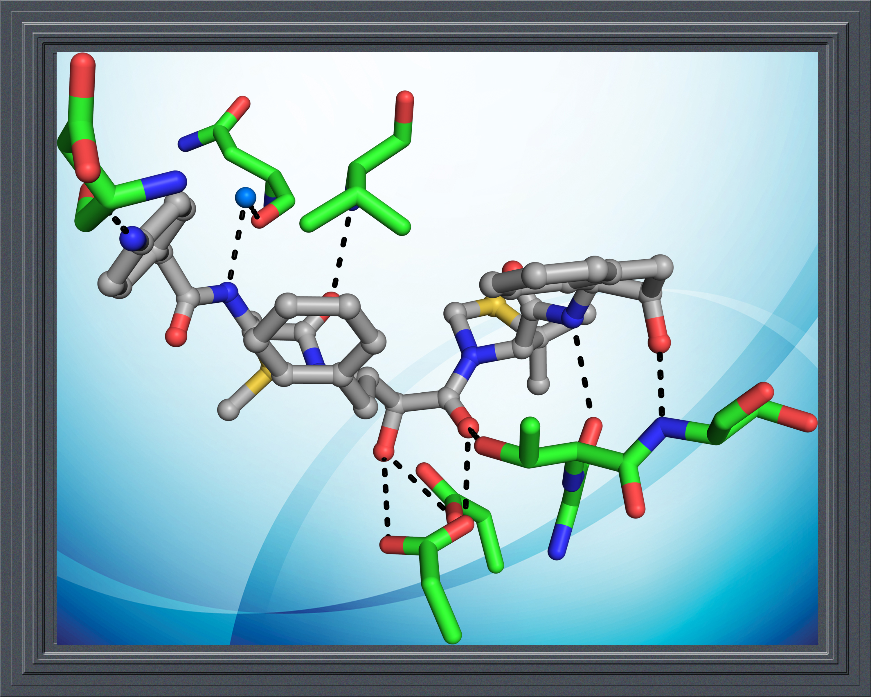
KNI-10343 bound crystal structure of plasmepsin II. PDB ID: 5YIA |
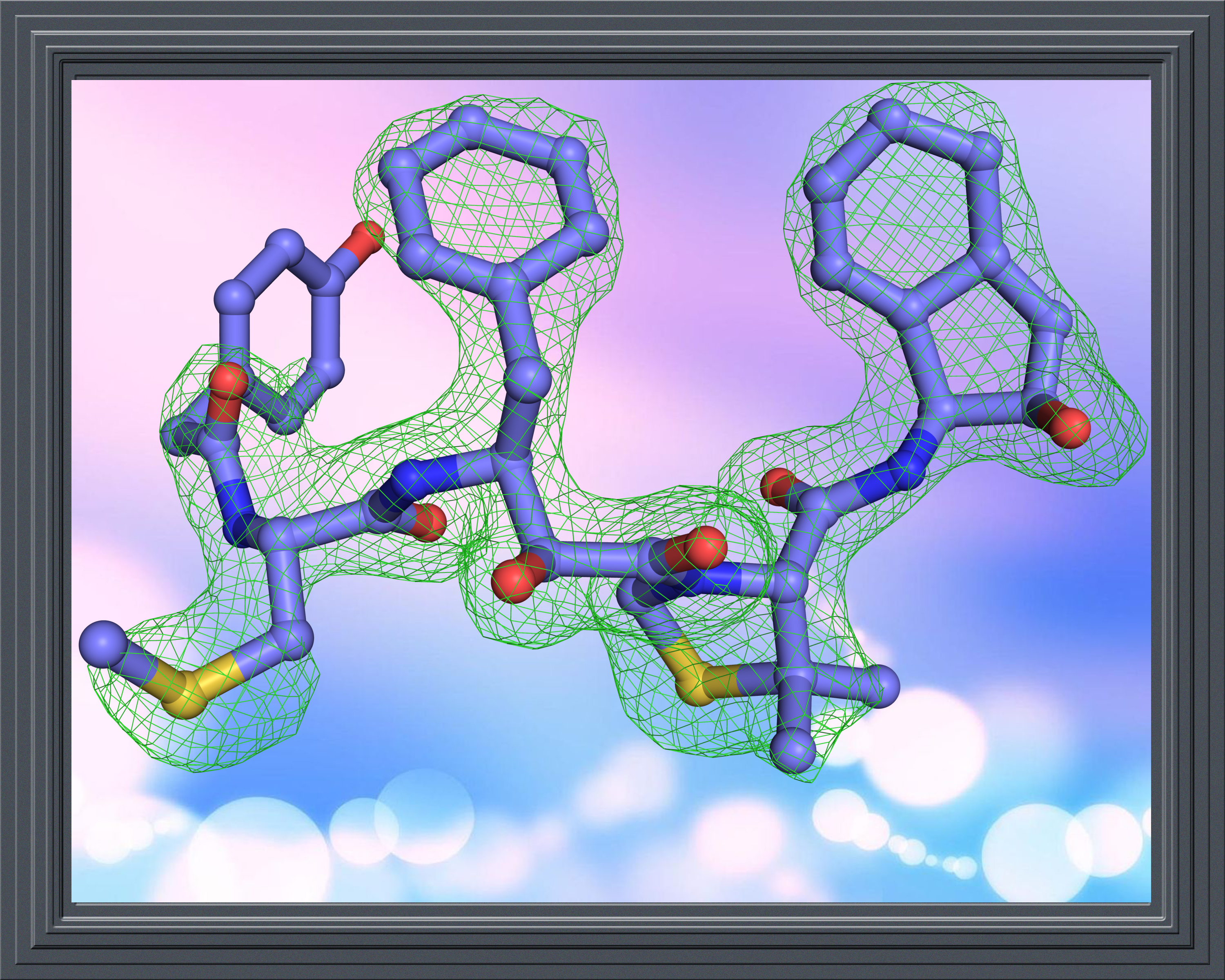
Crystal structure of plasmepsin II complexed with KNI-10395. PDB ID: 5YID |
|---|
 |
 |
 |
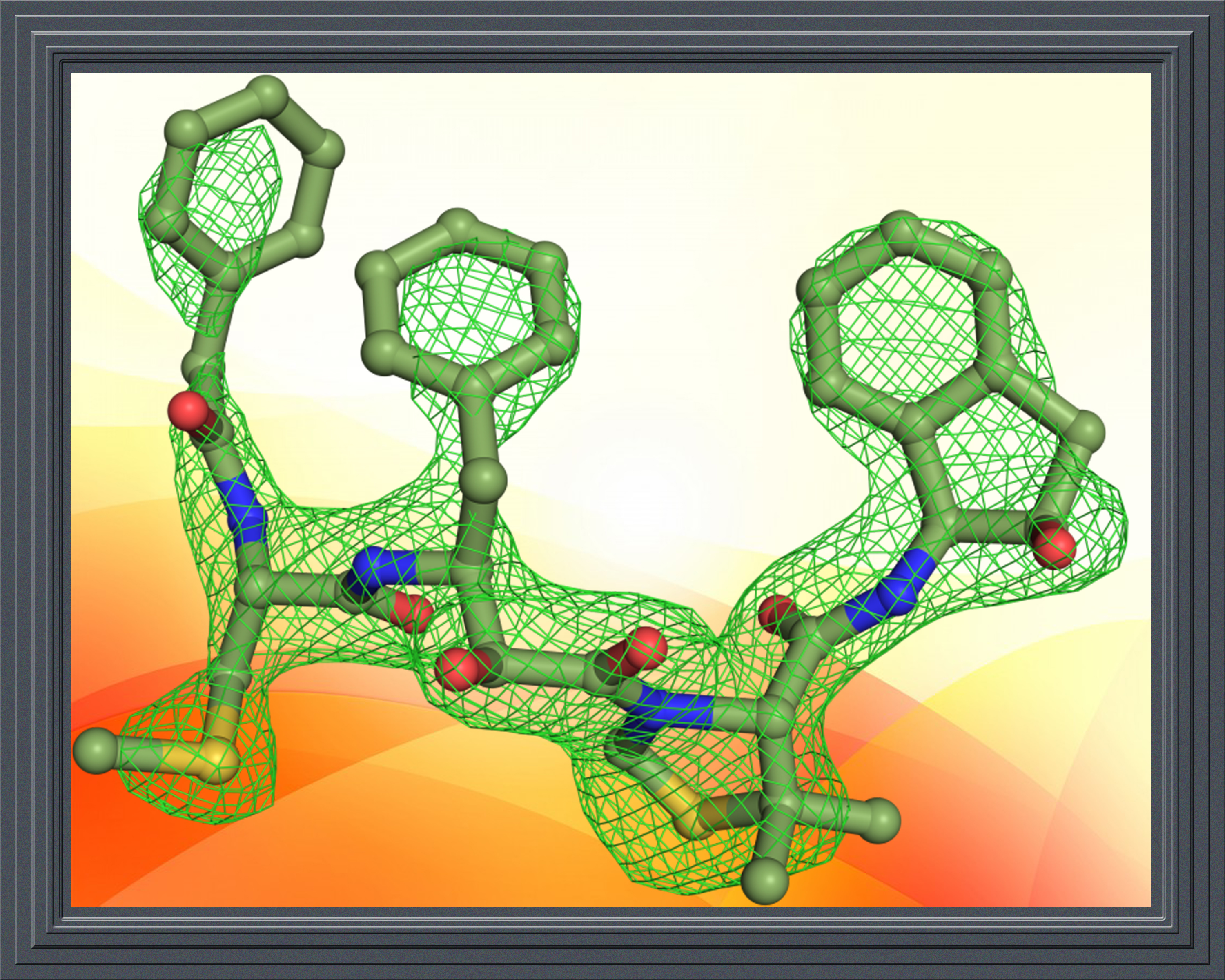
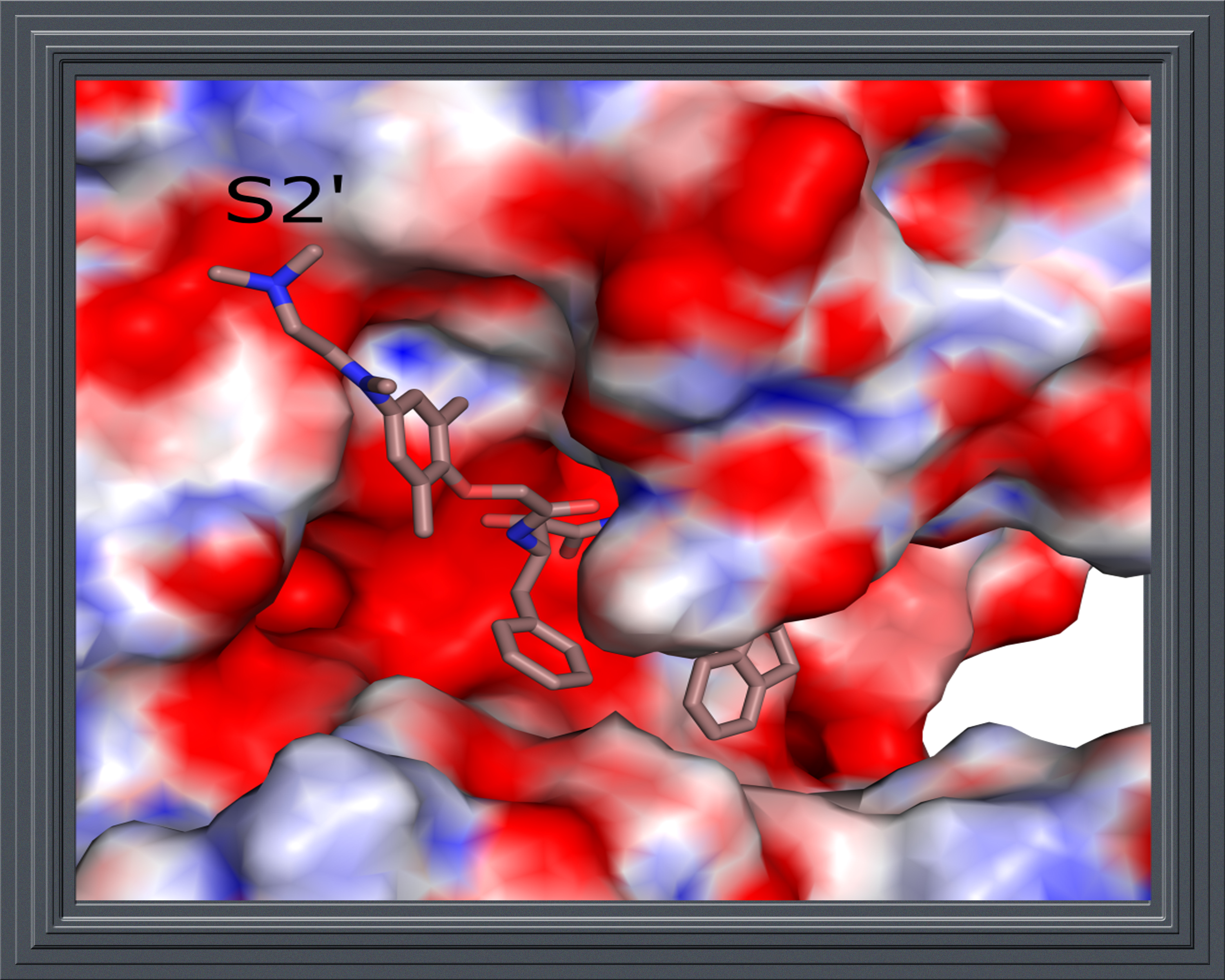
KNI-10742 complexed crystal structure of plasmepsin II. PDB ID: 5YIE |
Crystal structure of plasmepsin II complexed with KNI-10743. PDB ID: 5YIB |
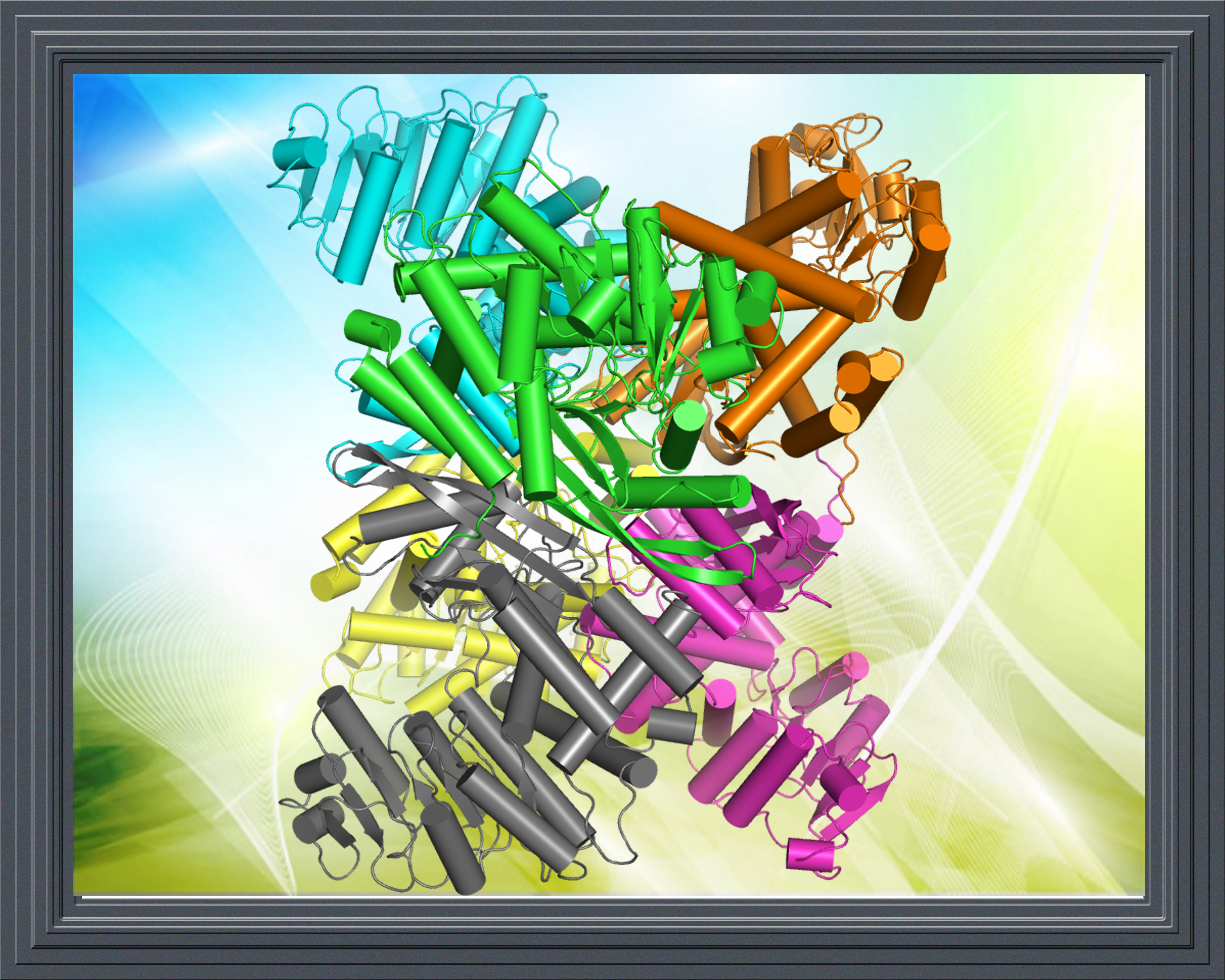
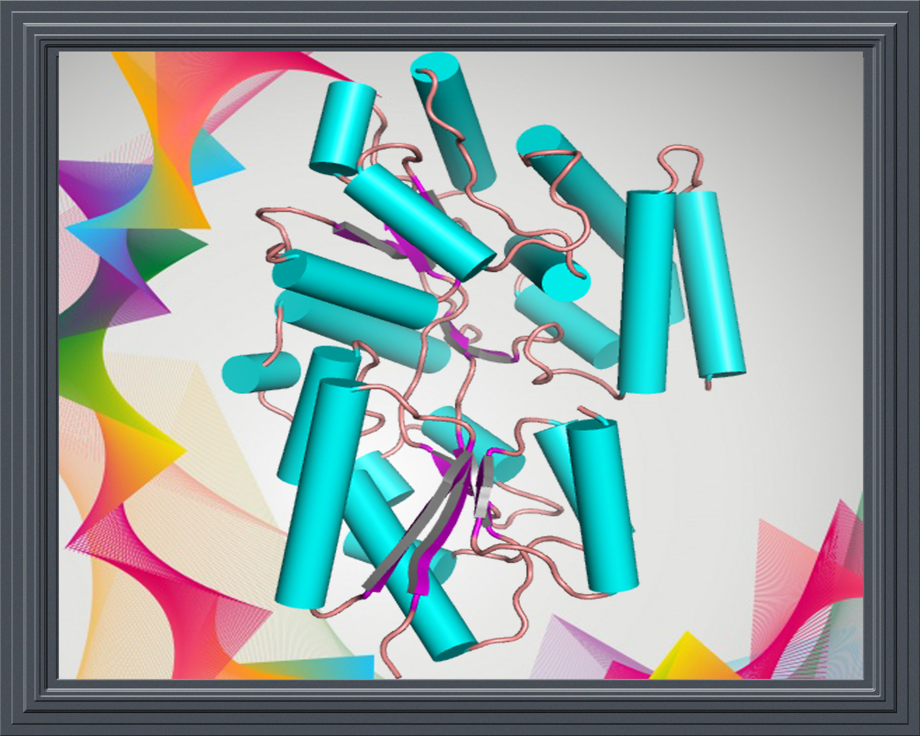
Crystal structure of A. niger Glutamate dehydrogenase (AnGDH) (apo form). PDB ID: 5XVI |
|---|
 |
 |
 |
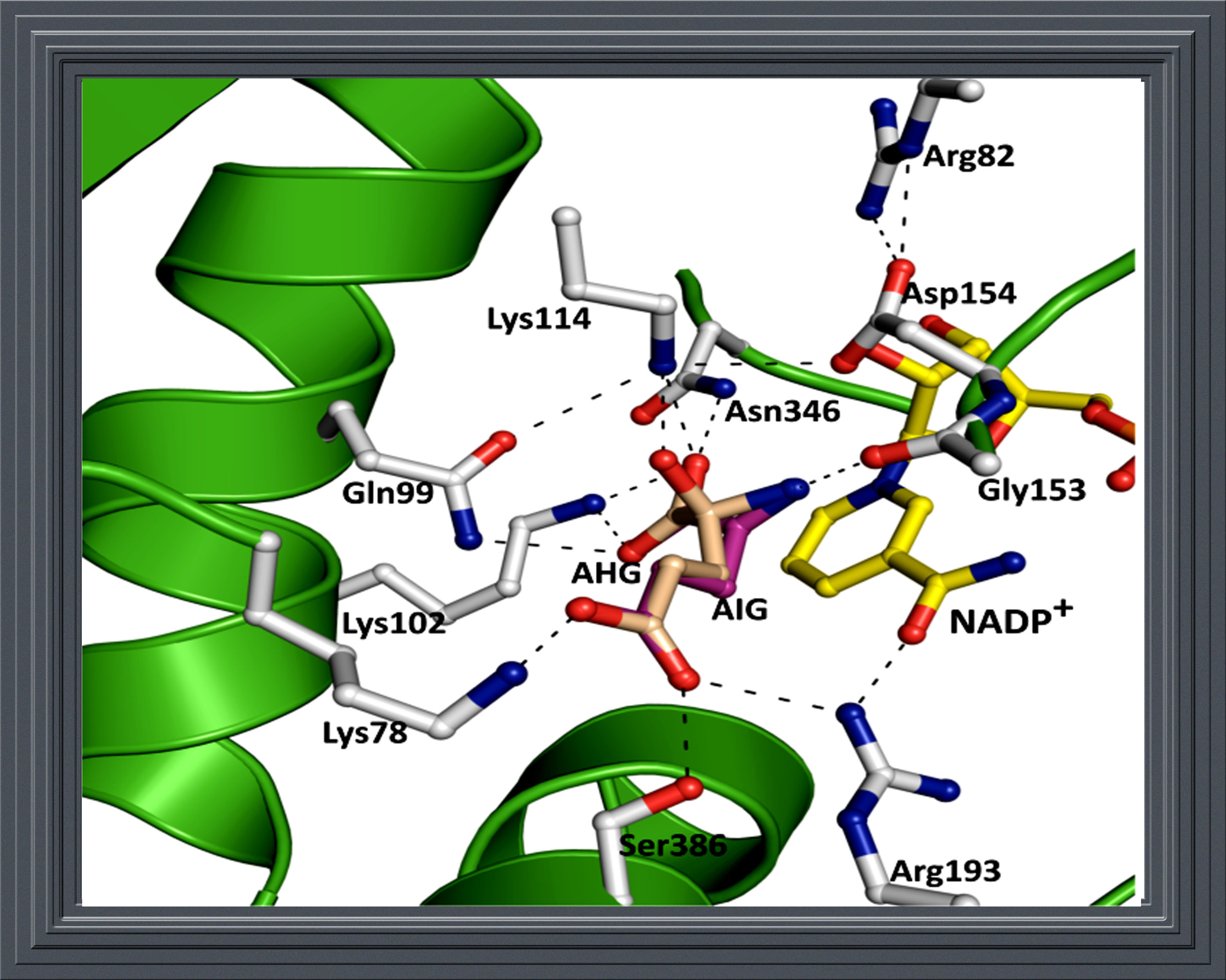
Crystal structure of A. niger Glutamate dehydrogenase (AnGDH) complexed with alpha-ketoglutarate (substrate) and NADPH. PDB ID: 5XVX |
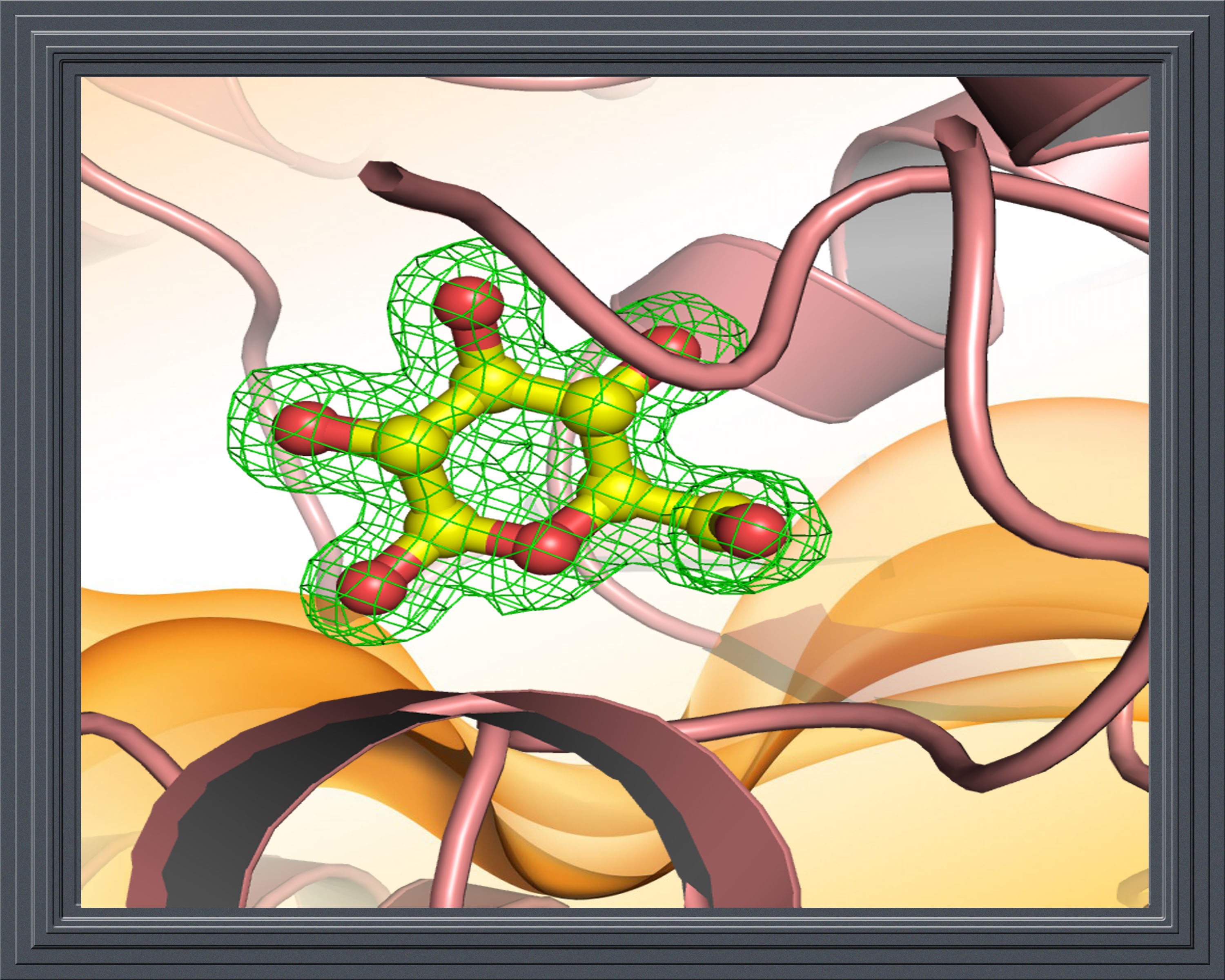
Crystal structure of A. niger Glutamate dehydrogenase (AnGDH) complexed with isophthalate (an inhibitor) and NADPH. PDB ID: 5XW0 |
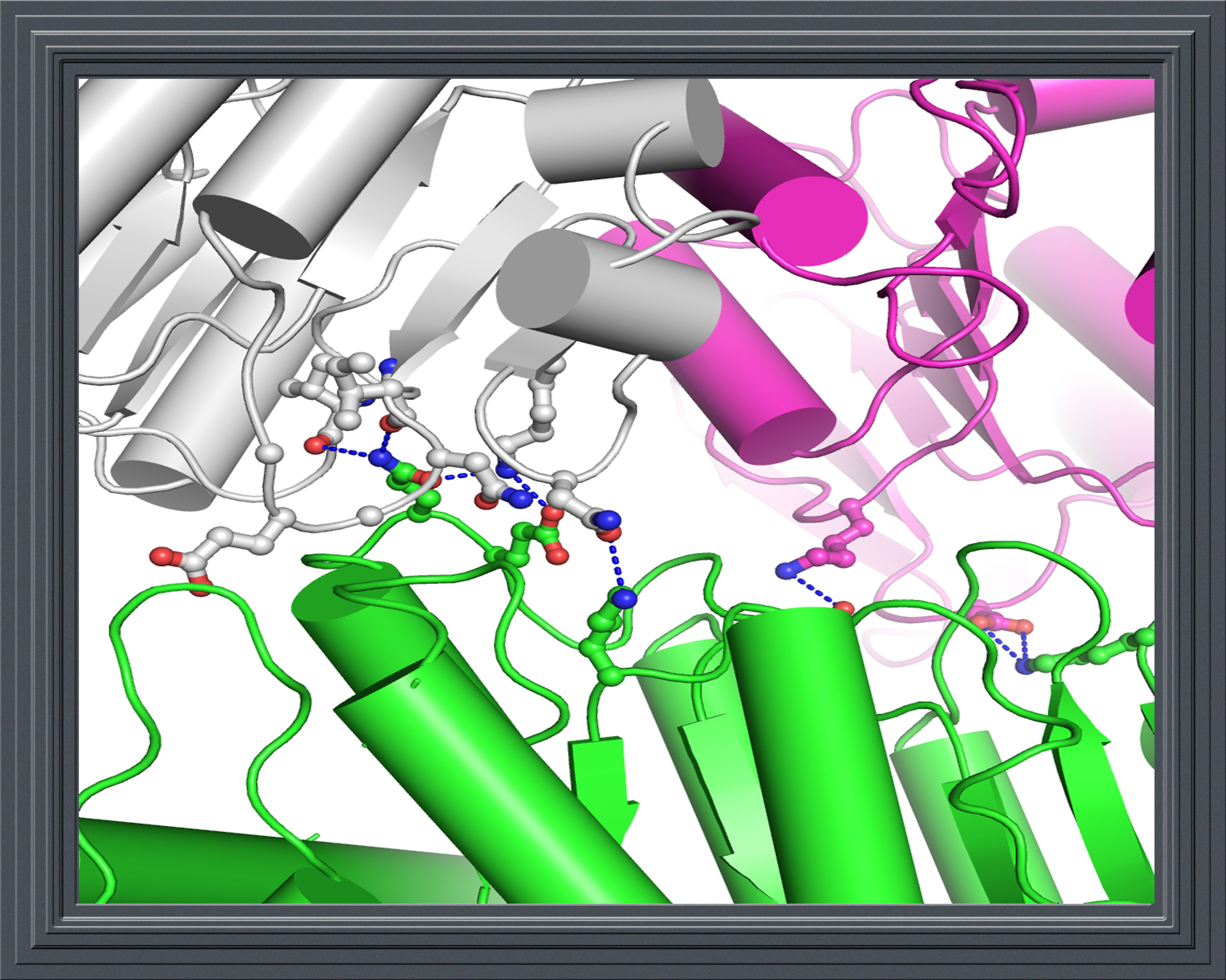
Forward inhibited form of A. niger Glutamate dehydrogenase (AnGDH) complexed with substrate alpha-ketoglutaratate in the active site. PDB ID: 5XVV |
|---|
 |
 |
 |
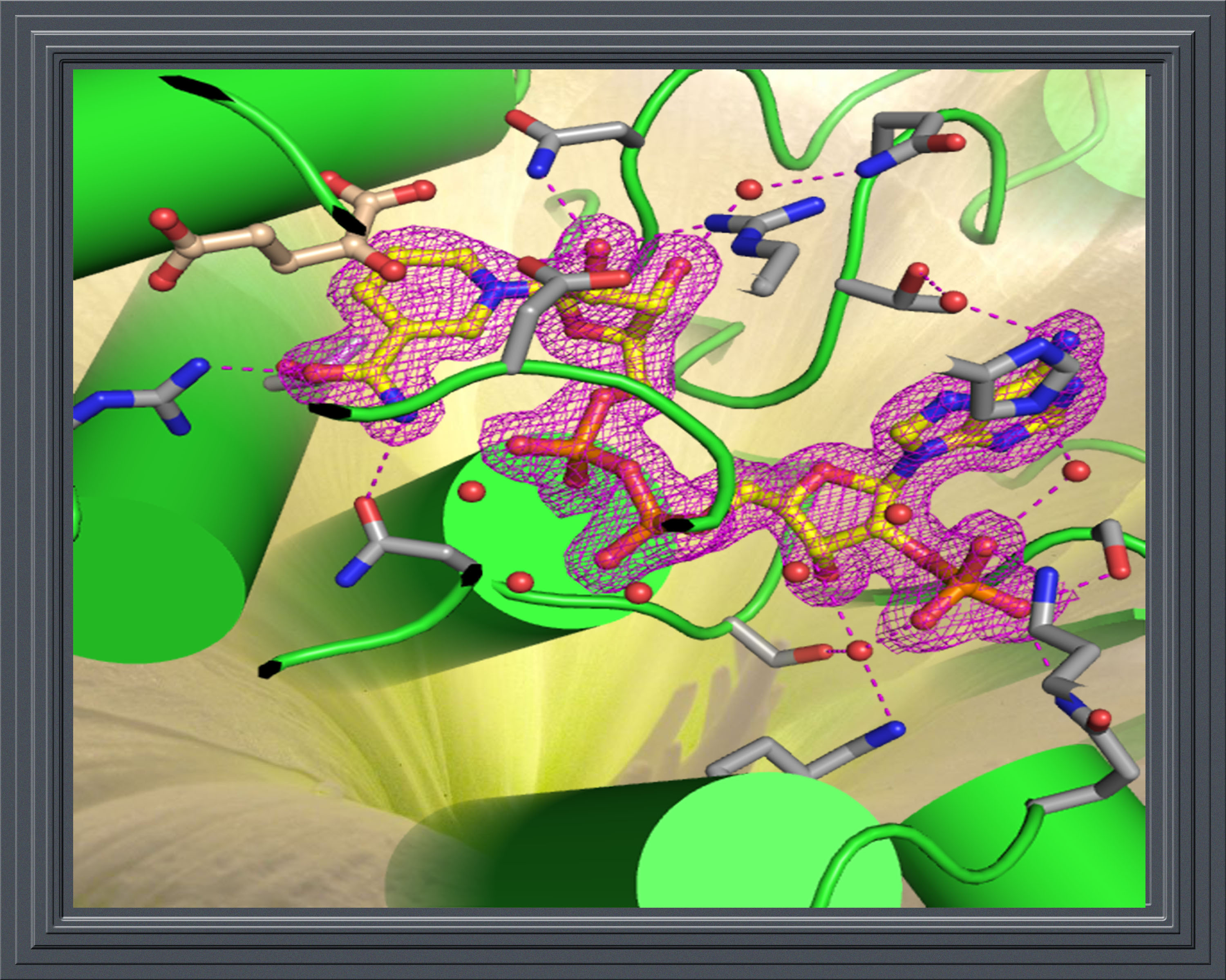
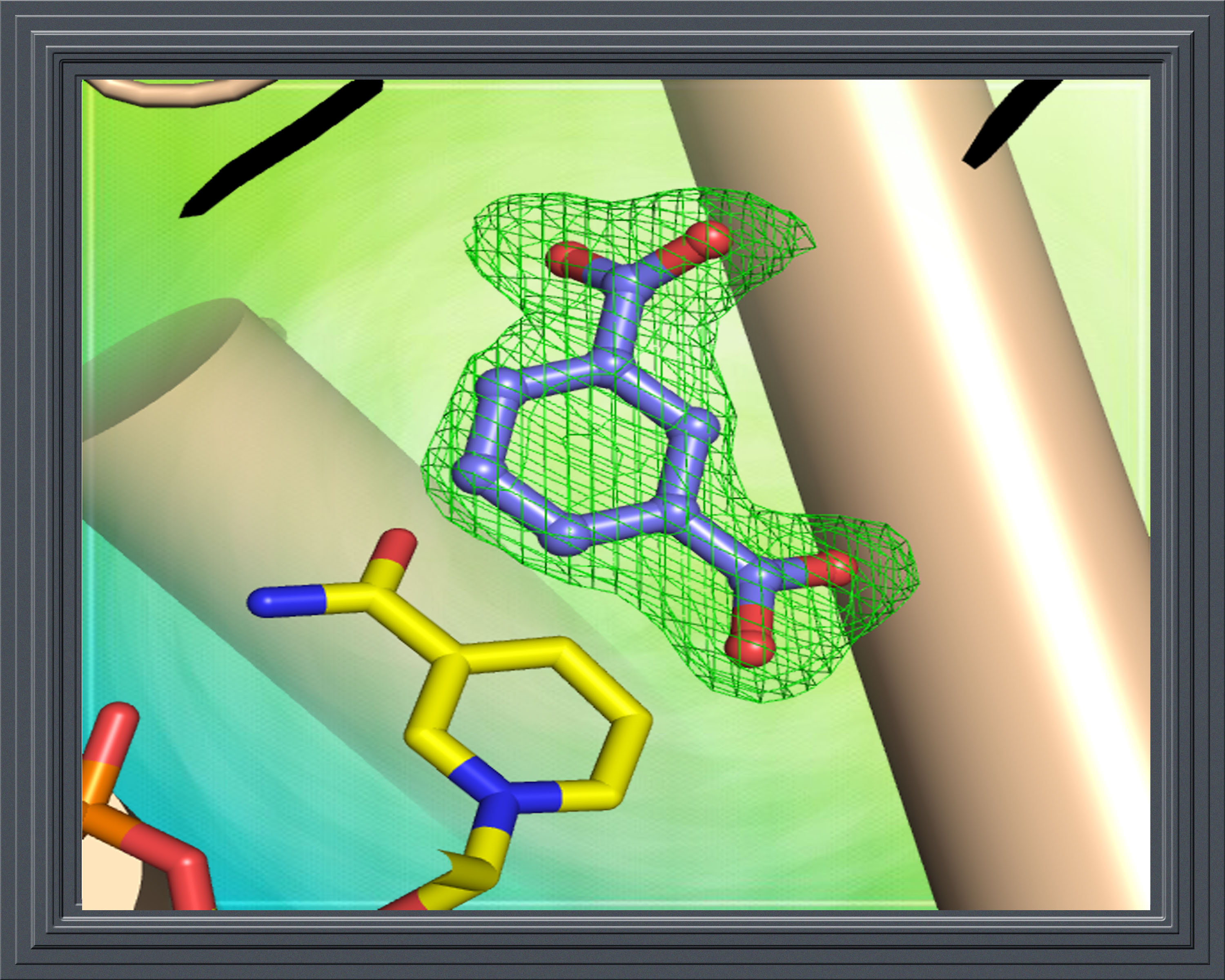
A. niger Glutamate dehydrogenase (AnGDH) structure bound with reaction intermediates. PDB ID: 5XWC |
Crystal structure of Pseudomonas putida glucose binding protein (apo-form). PDB ID: 5DVF |
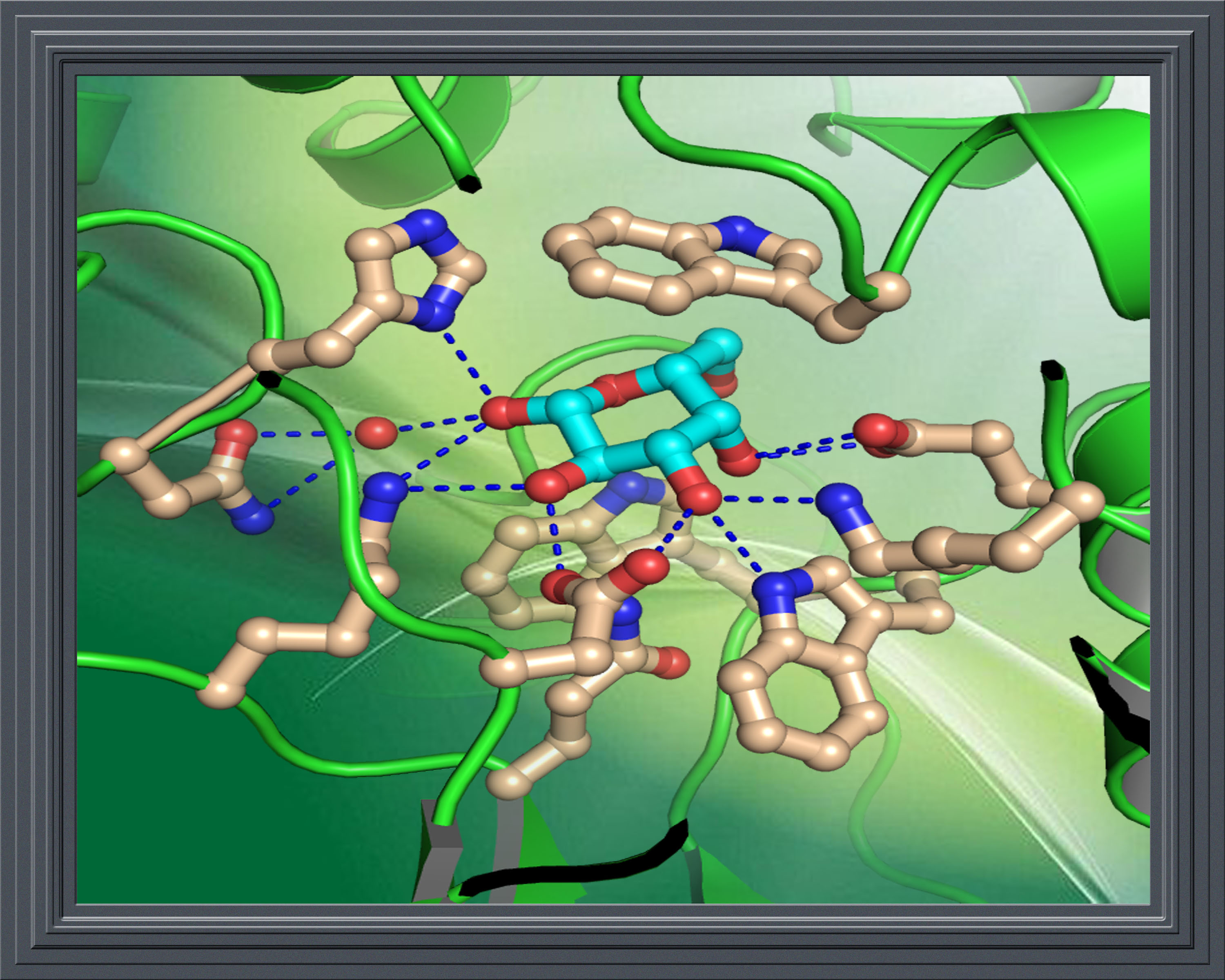
High resolution crystal structure of glucose complexed Pseudomonas putida glucose binding protein. PDB ID: 5DVI |
|---|
 |
 |
 |
Galactose bound form of Pseudomonas putida glucose binding protein. PDB ID: 5DVJ |
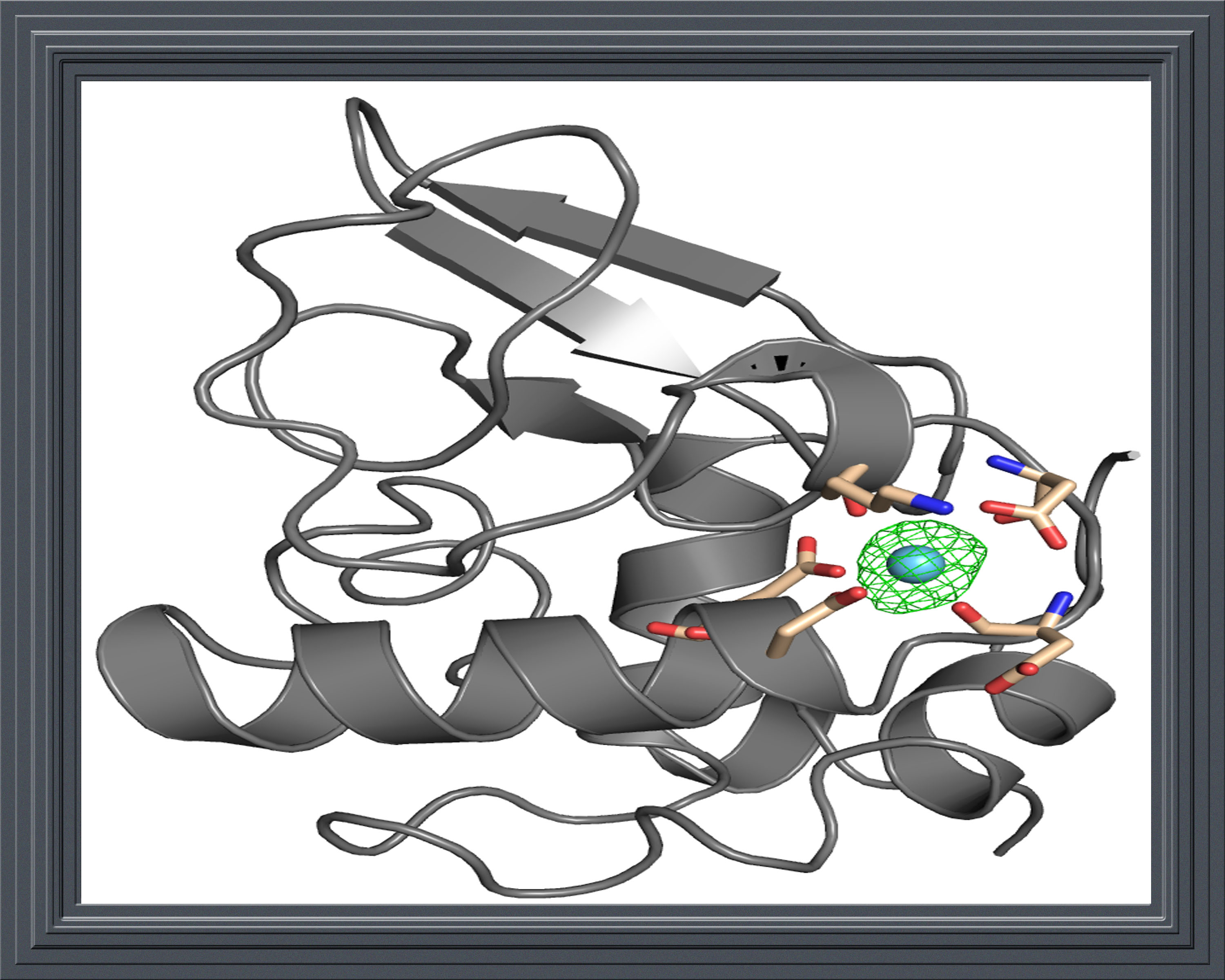
Crystal structure of Lanthanum ion bound bovine alpha-lactalbumin. PDB ID: 5X84 |
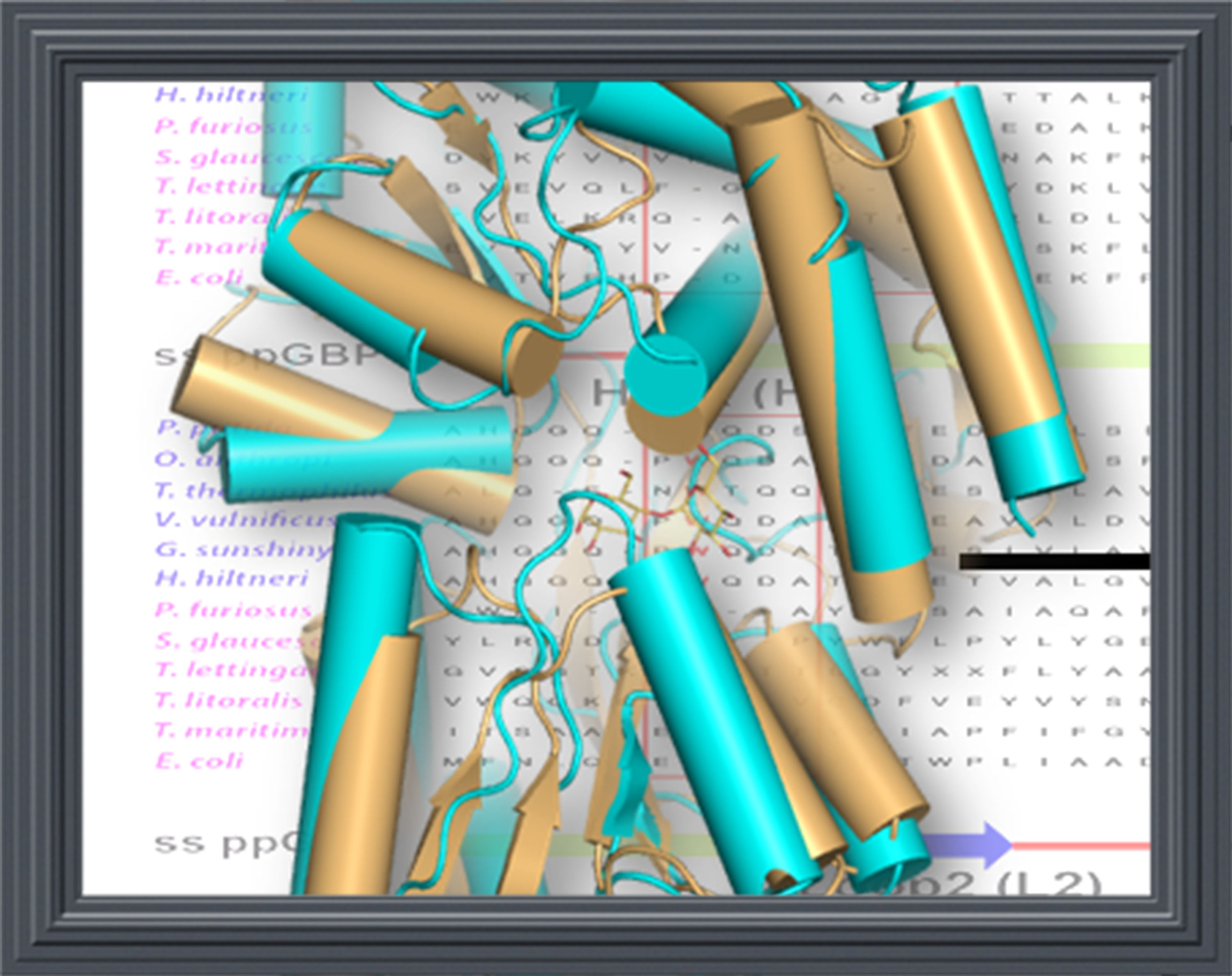
Crystal Structure of Periplasmic glucose binding protein ppGBP deletion mutant- Del-ppGBP. PDB ID: 5XPJ |
|---|